

Bile Acids: Which Sample Type Should I Study?
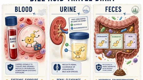
We’re often asked what matrices are compatible with our bile acids kit. That answer is easy (pretty much all of them). But the more relevant question is probably: What is the Best Sample? That answer is more complex, and the best sample might be the one that best answers your biological question, or simply the one that you have.
Why are we talking about the sample matrix for bile acids?
Bile acids move through a remarkably distributed system. They are synthesized and conjugated in the liver, released into the intestine, transformed by gut microbes, reabsorbed through enterohepatic circulation, exposed to tissues (including crossing the blood-brain barrier!) and are ultimately eliminated in feces or urine. A blood draw, urine cup and stool sample therefore do not offer three interchangeable views of the same pool, but each catches bile acids at a different point in that journey. [1,2]
That difference is likely meaningful. The table below is a quick summary of the most common matrices studied in bile acids research, along with why each is important, what it might be best suited for, and the main cautions or practical limitations for each. The practical question is not simply which matrix contains the most bile acids, but rather a two-part question of what part of bile acid biology you want to observe, and to which samples do you actually have access.
A quick matrix guide
| Matrix | What it emphasizes | Best suited for | Main caution | Coverage (MoveKit BA) | Most Abundant BA* |
|---|---|---|---|---|---|
| Serum / plasma | Systemic exposure and enterohepatic spillover | Cohorts, longitudinal response, liver/metabolic phenotypes | Meals, time of day, matrix | ~28 quantitative | GCDCA / CDCA |
| Urine | Renal clearance and phase-II conjugation | Sulfated/glucuronidated BAs, repeat sampling | Hydration, renal function, normalization | ~24 quantitative | GDCA-3S |
| Feces | Distal gut and microbial transformation | Microbiome function, primary-to-secondary conversion, MCBAs | Heterogeneity, transit, collection | 12-32 quantitative, highly variable | DCA |
| Tissue / CSF / cells | Local pools or controlled mechanisms | Transport, receptor biology, spatial questions | Invasiveness, low abundance, limited context | Varies | Varies |
Table 1. No matrix is universally superior; each emphasizes a different biological compartment and set of preanalytical risks. *Most abundant as detected in a pool of 400 normal human donors. Can vary widely by individual.
Blood: the practical view of systemic exposure
For most human studies, serum or plasma is the easiest place to begin. Blood collection is familiar, biobanks already contain samples, and volumes as low as 20-30 uL can support quantitative LC-MS analysis. Circulating bile acids have been shown to be useful for studying hepatobiliary dysfunction, metabolic and cardiovascular disease, inflammation, therapeutic response and the systemic side of the gut-liver-brain axis. [2,3]
The strength of blood is also its limitation: it is an integrated downstream signal. A change in circulating DCA, for example, could involve microbial production, intestinal absorption, hepatic reconjugation, transporter activity or renal/hepatobiliary clearance. Ratios of secondary-to-primary or conjugated-to-unconjugated bile acids can sharpen interpretation, while broader coverage of sulfated species and MCBAs can reveal remodeling that a total bile acid value conceals.
For cross-sectional biomarker studies, a standardized fasting collection at a consistent time is usually the cleanest design. For physiology studies, however, the response may be the signal: a meal challenge with serial samples can reveal bile acid kinetics that a single fasting draw misses. [2,4]
Does serum versus plasma make a meaningful difference?
Both are defensible, and the biological questions they answer are largely the same. Plasma avoids the clotting interval and is often preferred for standardized metabolomics workflows. However, the clinical testing community seems to have harmonized to using serum for clinical bile acids measurements, and serum may be more abundant in existing clinical collections. Values can vary by matrix, so choose one matrix and anticoagulant, document the protocol, and do not mix serum and plasma within a comparison unless the design explicitly tests that difference. A recent standardization review recommended fasting plasma as a practical default, while emphasizing that consistency across the entire workflow matters more than the label on the tube. [2,5]
Urine: a view of bile acids from one exit ramp
Urine is attractive because it is noninvasive, inexpensive and easy to collect repeatedly at home or in large cohorts. Biologically, it asks a more specific question than blood: which bile acids has the body slated for renal elimination? Sulfation and glucuronidation increase hydrophilicity and reduce intestinal reabsorption, creating an alternate clearance route when hydrophobic bile acids accumulate or hepatobiliary transport is stressed. In healthy subjects, one LC-MS/MS study found that sulfated species represented about 89% of urinary bile acids, compared with 33% of serum bile acids. Urine is therefore not merely dilute plasma; it strongly emphasizes phase-II conjugation and clearance. [6,7]
That emphasis makes urine particularly useful for studies of sulfated bile acids, cholestasis, drug-induced liver injury, inborn errors of bile acid metabolism and the gut-liver-kidney axis. Because collection is easy, urine also supports dense longitudinal sampling: before and after a meal, during a treatment course or across days. It may expose an adaptive detoxification response even when the total circulating pool changes only modestly.
As with all urine metabolite measurements, interpretation requires attention to dilution and kidney function. Creatinine normalization is common for spot samples, yet creatinine itself varies with muscle mass, diet, time of day and kidney function; specific gravity or osmolality may be useful complementary approaches. Whatever strategy is chosen should be prespecified and applied consistently. Renal impairment is especially important: a high serum sulfate with low urinary excretion could reflect reduced clearance rather than increased bile acid production. [8]
Feces: the closest practical look at microbial bile acid chemistry
If the primary question concerns the gut microbiome, feces often provides the most direct practical matrix. Most bile acids are reabsorbed before they leave the body, but the residual fecal pool has traveled through the distal intestine, where microbial bile salt hydrolases deconjugate host bile acids and other enzymes drive oxidation, epimerization and 7α-dehydroxylation. Fecal profiles can therefore reveal the balance between primary and secondary bile acids, the extent of deconjugation and the appearance of microbial products that may be scarce or absent in peripheral blood. Pairing these measurements with metagenomics helps connect “who is there?” with “what chemistry are they performing?” [1,9,10]
Feces may be especially informative for diet interventions, antibiotics, probiotics, inflammatory bowel disease, colorectal cancer and other studies where intestinal ecology is central. It is also a logical discovery matrix for MCBAs because these compounds are formed in the gut and may not reach high systemic concentrations. But fecal abundance is not synonymous with systemic exposure. A bile acid can be abundant in stool because it was poorly absorbed, rapidly produced or simply concentrated by water loss. Conversely, a biologically potent species could be efficiently reabsorbed and leave only a small fecal trace. Plasma and feces measurements can answer complementary questions.
The tradeoff is practical, and mostly to do with collection and processing. Stool is heterogeneous, varies markedly in water content, and is influenced by diet, transit time and collection method. Representative sampling requires either thorough homogenization or a defined multisite subsampling strategy. Investigators must also decide whether to normalize to wet weight, dry weight, protein normalization or another measure. In one study of 70 human samples, wet-weight normalization produced the narrowest distribution, but the broader lesson is that normalization choice changes the apparent quantitative result and must remain consistent. [11,12]
Less standardized than clinical blood draws or urine sampling, collection matters for feces. Immediate freezing is a strong default when feasible, to quench enzyme and microbe activity, but often impractical. A study comparing stabilized and snap-frozen stool found reasonable agreement for relative patterns of common bile acids but significantly different total concentrations; the two methods should not be mixed without a bridging study. Feces offers unusually rich biology, and rewards careful logistics. [13]
Less common sample types: more specific questions, more specialized studies
CSF and brain tissue
Cerebrospinal fluid can bring a study closer to the central nervous system than peripheral blood, which is appealing for neurodegeneration and gut-brain-axis research. The tradeoffs are invasiveness, limited volume and very low concentrations for many bile acids. CSF profiles may reflect transport across the blood-brain barrier, local cholesterol metabolism or disease-related barrier changes, but they should not automatically be interpreted as diluted plasma. Postmortem brain tissue adds spatial and pathway information and has supported associations between bile acid networks and Alzheimer’s disease, although agonal state, postmortem interval and terminal disease complicate interpretation. [14,15]
Liver, intestinal tissue, bile and luminal contents
Tissue and bile place the measurement close to synthesis, transport and local receptor exposure. Liver tissue can be valuable for cholestasis, transporter biology and drug-safety studies; intestinal tissue can connect luminal chemistry with epithelial or immune responses. Bile aspirates and ileal or cecal contents offer a more proximal and spatially resolved view than feces, often making them especially informative in animal models or surgical studies. They are correspondingly invasive, less scalable and more sensitive to anatomical sampling location. A fecal sample is an endpoint of the gut journey; an ileal or cecal sample can reveal where a transformation occurred. [9,16]
Cells, microbial cultures and organoids
Controlled systems are best when the goal is mechanism. Hepatocytes can test synthesis, sulfation or transporter inhibition; bacterial cultures can establish whether a strain converts a defined substrate; intestinal or liver organoids can probe receptor signaling and tissue-to-tissue interactions. Measuring both cells and conditioned medium distinguishes intracellular accumulation from secretion or uptake. These models simplify causal inference, but they deliberately remove the integrated physiology that makes bile acid biology complex. Medium composition, serum supplements and bile acid adsorption also require matrix-matched controls. [17,18]
Dried blood spots and microsampling
Dried blood spots or volumetric microsamples may be useful when repeated venipuncture, remote collection or pediatric sampling is impractical. They can make longitudinal designs much easier, but low sample volume, hematocrit effects and analyte-specific recovery must be validated. They are a logistical innovation, not yet a drop-in replacement for plasma. [19]
Choose the simplest matrix that can answer the biological question
Convenience is not a scientific compromise when the matrix is aligned with the hypothesis. Serum or plasma is often the best first choice for scalable human phenotyping and systemic signaling. Urine is exceptionally useful when phase-II conjugation and renal elimination are central. Feces earns its additional processing burden when the microbiome and intestinal transformations are the main event. Specialized matrices become worthwhile when local exposure or mechanism cannot be inferred from an accessible biofluid.
When resources permit, paired matrices can be more informative than simply expanding cohort size in one compartment. Plasma plus feces separates systemic exposure from distal gut chemistry. Serum or plasma plus urine helps distinguish circulating accumulation from renal clearance. Tissue plus blood connects local mechanism to a translational biomarker. The most revealing design may be the one that samples two well-chosen points along the same bile acid journey.
MoveKit™ BA supports broad bile acid profiling across serum/plasma, urine, feces and selected tissues, combining quantitative coverage of established bile acids - including sulfated species - with semiquantitative coverage of emerging chemistry such as microbial-conjugated bile acids. The purpose of a flexible workflow is not to make every matrix equivalent. It is to let the biology determine where you look.
References
- Fleishman JS, et al. Bile acid metabolism and signaling in health and disease: molecular mechanisms and therapeutic targets. Signal Transduct Target Ther. 2024;9:97. Link
- Joseph S, et al. Towards the clinical use of peripheral bile acids: recommendations to limit their preanalytical and analytical sources of variability. Pract Lab Med. 2025;47:e00508. Link
- MahmoudianDehkordi S, et al. Altered bile acid profile associates with cognitive impairment in Alzheimer’s disease - an emerging role for gut microbiome. Alzheimers Dement. 2019;15:76-92. Link
- Lemaziere A, et al. Distinct postprandial bile acid responses to a high-calorie diet in men with normal and impaired glucose metabolism. 2020. Link
- Thachil A, et al. An overview of pre-analytical factors impacting metabolomics analyses of blood samples. 2024. Link
- Bathena SPR, et al. The profile of bile acids and their sulfate metabolites in human urine and serum. J Chromatogr B. 2013;942-943:53-62. Link
- Perreault M, et al. Urinary elimination of bile acid glucuronides under severe cholestatic conditions in humans. 2018. Link
- Tang KWA, et al. Normalisation of urinary biomarkers to creatinine for clinical practice and research - when and why. 2015. Link
- Wegner K, et al. Rapid analysis of bile acids in different biological matrices using LC-ESI-MS/MS for investigation of bile acid transformation by mammalian gut bacteria. Anal Bioanal Chem. 2017;409:1231-1245. Link
- Marion S, et al. Biogeography of microbial bile acid transformations along the murine gut. J Lipid Res. 2020;61:1450-1463. Link
- Schott HF, et al. Evaluation of normalization approaches for quantitative analysis of bile acids in human feces. Metabolites. 2022;12:723. Link
- Hu C, et al. Heterogeneity and lyophilization comparison of stool processing for gastrointestinal bile acid measurement by LC-MS/MS. J Chromatogr B. 2023. Link
- Neuberger-Castillo L, et al. Fitness for purpose of stabilized stool samples for bile acid metabolite analyses. Sci Rep. 2021;11:7904. Link
- Ogundare M, et al. Cerebrospinal fluid steroidomics: are bioactive bile acids present in brain? J Biol Chem. 2010;285:4666-4679. Link
- Baloni P, et al. Metabolic network analysis reveals altered bile acid synthesis and metabolism in Alzheimer’s disease. Cell Rep Med. 2020;1:100138. Link
- Shalon D, et al. Profiling the human intestinal environment under physiological conditions. Nature. 2023;617:581-591. Link
- Dewaele D, et al. LC-MS/MS analysis of bile acids in in vitro samples. Methods Mol Biol. 2019;1985:67-80. Link
- Blutt SE, et al. Use of human tissue stem cell-derived organoid cultures to model enterohepatic circulation. Am J Physiol Gastrointest Liver Physiol. 2021. Link
- Engevik MA, et al. Repurposing dried blood spot device technology to examine bile acid profiles. Sci Rep. 2024;14:15167. Link
About the Author
J. Will Thompson is a Founding Partner and serves as Operations Lead at Move Analytical LLC.